
So, what about wellbeing?
One of the most preventable workplace illnesses is hearing loss. The hazard that we don’t see but hear is noise.
Overview
The Centers for Disease Control (CDC) estimates that 22 million workers are exposed to potentially damaging noise at work each year. Whether you work at a sports venue, on a tarmac, or operate a jackhammer, hearing loss is preventable.
Know Your Workplace Noise Levels!
If you need to raise your voice to speak to someone 3 feet away, noise levels might be over 85 decibels. Several sound-measuring instruments are available to measure the noise levels in a workspace. These include sound level meters, noise dosimeters, and octave band analyzers.
Noise may be a problem in your workplace if you:
- Hear ringing or humming in your ears when you leave work.
- Must shout to be heard by a coworker an arm’s length away.
- Experience temporary hearing loss when leaving work.
The National Institute for Occupational Safety and Health (NIOSH) Sound Level Meter App is one tool available to the public to download on mobile iOS devices that measures sound levels in the workplace and provides noise exposure parameters to help reduce occupational noise-induced hearing loss. NIOSH Sound Level Meter on the App Store
Health Effects
Exposure to high levels of noise can cause permanent hearing loss. Neither surgery nor a hearing aid can help correct this type of hearing loss. Short-term exposure to loud noise can also cause a temporary change in hearing (your ears may feel stuffed up) or a ringing in your ears (tinnitus). These short-term problems may go away within a few minutes or hours after leaving the noise. However, repeated exposures to loud noise can lead to permanent tinnitus and/or hearing loss.
Loud noise can create physical and psychological stress, reduce productivity, interfere with communication and concentration, and contribute to workplace accidents and injuries by making it difficult to hear warning signals. The effects of noise-induced hearing loss can be profound, limiting your ability to hear high-frequency sounds, understand speech, and seriously impairing your ability to communicate.
How does the ear work?
When sound waves enter the outer ear, the vibrations impact the ear drum and are transmitted to the middle and inner ear. In the middle ear three small bones called the malleus (or hammer), the incus (or anvil), and the stapes (or stirrup) amplify and transmit the vibrations generated by the sound to the inner ear. The inner ear contains a snail-like structure called the cochlea which is filled with fluid and lined with cells with very fine hairs. These microscopic hairs move with the vibrations and convert the sound waves into nerve impulses–the result is the sound we hear.
Exposure to loud noise can destroy these hair cells and cause hearing loss!
What do I need to know about the anatomy and physiology of the ear?
The ear is the organ that makes hearing possible. It can be divided into three sections:
- The external outer ear
- The air-filled middle ear
- The fluid-filled inner ear
Outer Ear
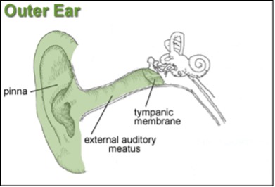
The parts of the outer ear include:
Pinna
- The pinna is the visible portion that is generally referred to as “the ear.”
- Its function is to localize sound sources and direct sound into the ear.
- The dimensions and folds of the pinna cause certain sound frequencies to be amplified and other frequencies to be weakened.
- Each individual’s pinna puts a distinctive imprint on the acoustic wave traveling into the auditory canal.
External Auditory Meatus (ear canal)
- The ear canal extends from the pinna to the eardrum and is about 26 millimeters (mm) in length and 7 mm in diameter. Size and shape vary among individuals. This is an important factor to consider when fitting hearing protectors.
- The ear canal protects the eardrum and acts as a resonator, providing about 10 decibels (dB) of gain to the eardrum at around 3,300 Hertz (Hz).
- The net effect of the head, pinna, and ear canal is that sounds in the 2,000 to 4,000 Hz region are amplified by 10 to 15 dB.
- Sensitivity to sounds is greatest in this frequency region, and noises in this range are the most hazardous to the ability to hear.
Tympanic Membrane (eardrum)
- The eardrum separates the outer ear from the middle ear, creating a barrier that protects the middle and inner areas from foreign objects. It is somewhat cone-shaped in appearance and is about 17.5 mm in diameter.
- The eardrum vibrates in response to the sound pressure waves. The actual distance that the membrane moves is incredibly small (as little as one-billionth of a centimeter).
Middle Ear
The purpose of the middle ear is to conduct sound from the outer ear to the inner ear. There are three main features of the middle ear:
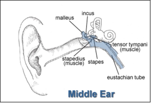
Ossicles (bones). The malleus (hammer), incus (anvil), and stapes (stirrup) make up the ossicles.
- The primary function of the middle ear is to transform the vibrating motion of the eardrum into the motion of the stapes. The middle ear enhances the transfer of this acoustical energy in two ways:
- The area of the eardrum is about 17 times larger than the oval window (see inner ear). The effective pressure (force per unit area) is increased by this amount.
- The ossicles produce a lever action that further amplifies the pressure. As a result, most of the energy entering normal ears through the eardrum is transmitted into motion of the stapes and stimulation of the inner ear system.
- Without the transformer action of the middle ear, only about 1/1000 of the acoustic energy in air would be transmitted to the inner-ear fluids (a loss of about 30 dB).
- The malleus and the incus vibrate together, transmitting the sound waves from the eardrum to the footplate of the stapes (this pushes the oval window in and out).
Muscles.
These include the tensor tympani and the stapedius.
- Attached to the malleus and stapes, the stapedius and tensor tympani muscles help keep the ossicles in their correct position and protect the internal ear from excessive sound levels.
- When the ear is exposed to sound levels above 80 dB, the muscles contract, decreasing the amount of energy transferred to the oval window.
- This protective reflex, known as the “aural reflex,” does not actually react fast enough to provide protection against impulse sounds, and the muscles do not stay contracted long enough to provide protection from long-term steady exposure.
Eustachian Tube.
- The eustachian tube connects the front wall of the middle ear with the nasal air passages.
- The eustachian tube also operates like a valve, which opens during swallowing.
- This equalizes the pressure on either side of the eardrum, which is necessary for optimal hearing. Without this function, a difference between the static pressure in the middle ear and the outside pressure may develop, causing the eardrum to displace inward or outward. This reduces the efficiency of the middle ear, and less acoustic energy will be transmitted to the inner ear.
Inner Ear
The purpose of the inner ear is to convert mechanical sound waves to neural impulses that can be recognized by the brain. The sensory receptors that are responsible for the initiation of neural impulses in the auditory nerve are contained in the cochlea of the inner ear.
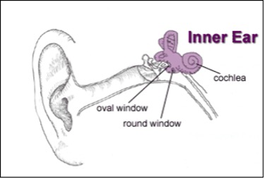
- The cochlea resembles a snail shell and spirals for about 2 3/4 turns around a bony column.
- Within the cochlea, there are three canals. They are called:
- The Scala VestibuliThe Scala Tympani (a bony shelf, called the spiral lamina, along with the basilar membrane and the spiral ligament, separates the upper Scala Vestibuli from the lower Scala Tympani)
- The Scala Media (cochlear duct)
- The Scala media is a triangular-shaped duct that contains the organ of hearing, called the “organ of Corti.”The basilar membrane, narrowest and stiffest near the oval window and widest at the tip of the cochlea, helps form the floor of the cochlear duct.
- The cochlear duct is separated from the Scala Vestibuli by Reissner’s membrane.
Hair Cells and Cilia
- The surface of the basilar membrane contains phalangeal cells that support the critical hair cells of the organ of Corti.
- The hair cells are arranged with an inner row of about 3,500 hair cells and three to five rows of approximately 12,000 outer hair cells.
- The cilia of the hair cells extend along the entire length of the cochlear duct and are embedded in the undersurface of the tectorial membrane.
- In general, the hair cells at the base of the cochlea respond to high-frequency sounds, while those at the apex respond to low-frequency sounds.
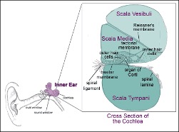
Activity in the Cochlea
- The movement of the stapedial footplate in and out of the oval window moves the fluid in the scala vestibuli.
- This fluid pulse travels up the Scala Vestibuli but causes a downward shift of the cochlear duct, along with distortion of Reissner’s membrane and a displacement of the organ of Corti.
- The activity is then transferred through the basilar membrane to the scala tympani.
- At the end of the cochlea, the round window acts as a relief point and bulges outward when the oval window is pushed inward.
- The vibration of the basilar membrane causes a pull, or shearing force, of the hair cells against the tectorial membrane.
- This bending of the hair cells activates the neural endings so that sound is transformed into an electrochemical response.
- This response travels through the Vestibulocochlear nerve, and the brain interprets the signal as sound.
Hearing loss in the workplace is preventable with the targeted training and education of our workforce. Do you hear me?